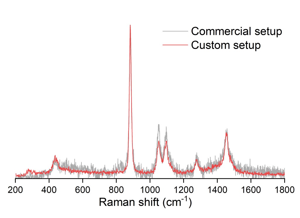
Raman microspectroscopic setup
In a currently running project, I have been constructing a new spectrometer for near-infrared Raman spectroscopy. As Raman scattering is an inherently weak process, highly sensitive detection is needed in the spectral range of interest. However, most near-infrared Raman setups still rely on costly detectors, which significantly reduce the applicability of this method. Meanwhile, using near-infrared excitation (e.g., at 785 nm) reduces sample fluorescence, which is a particularly attractive feature in biophysical applications. With the new spectrometer, the sensitivity is improved to an extent that allows to build it into a custom Raman microscope.
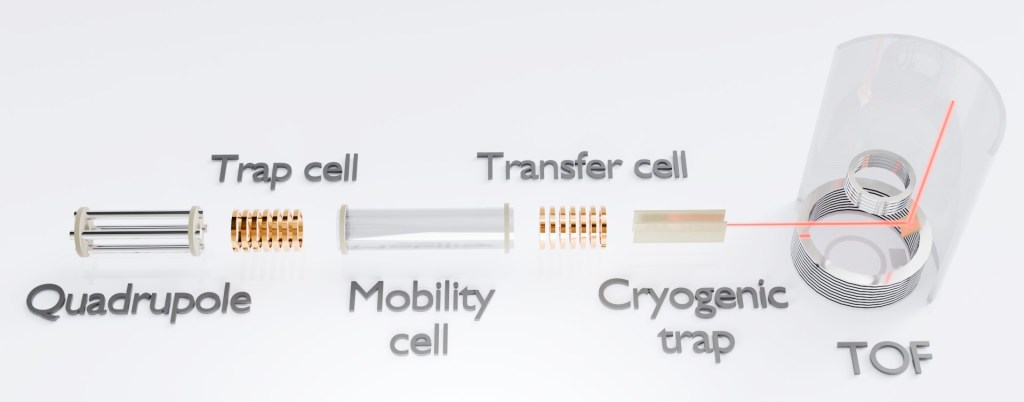
Gas-phase infrared action spectroscopy setup
Infrared spectroscopy relies on the absorption of infrared photons and the detection of absorbance or transmittance of the excitation light. While this is routinely done in condensed phase nowadays, its gas-phase application is seriously hindered by the fact that it scales with concentration. In a new setup that I have been developing for the past few years, we do infrared spectroscopy in a mass spectrometer! While this allows for highly fundamental insight into our molecules of interest, in a Coulomb-limited ion cloud, the analyte density is orders of magnitude lower than the detection threshold. Therefore, we employ messenger tagging spectroscopy. The ions are trapped in a potential well and cooled down to ~40 K. A small amount of N2 gas is introduced into the trap chamber, which precipitates on the ions due to non-specific interactions with the charges. This results in a mass shift of 28 Da-equivalent in a time-of-flight detector. The ions are then overlapped with tunable narrowband mid-infrared pulses. When resonant photons are absorbed, the additional energy of ions is dissipated by the depletion of N2 tags. By following the signal intensity of the tagged ion in fuction of the wavelength, we can recover the infrared spectrum with exceptional spectral resolution. The instrument was connected to both a free electron laser light source, as well as a benchtop pulsed OPO-OPA system.
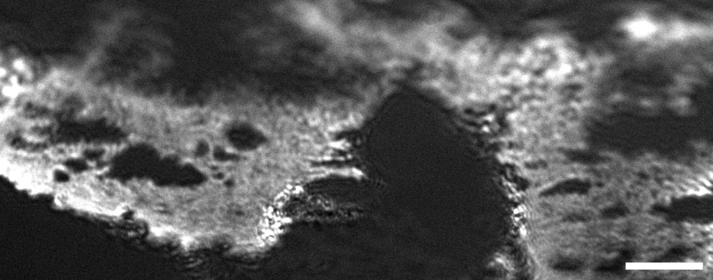
Brewster-angle microscope
Brewster-angle microscopy is a special case of polarization microscopy. When an interface is illuminated at its Brewster angle, only the s-polarized light is reflected from it. When thin films, such as lipid monolayers, are deposited at the air-water interface, it changes the refraction index of the surface, which changes the corresponding Brewster angle as well. Thus, where lipid rafts are present in the area illuminated with p-polarized light, high-contrast microscopic visualization is possible of the film morphology. With a high-framerate camera and near-infrared illumination, the interaction of lipid monolayers and their conjugates with relevant molecules of interest can be monitored in real time. As near-infrared illumination is used, parasite processes such as fluorescence of strong scattering do not reduce the image contrast.